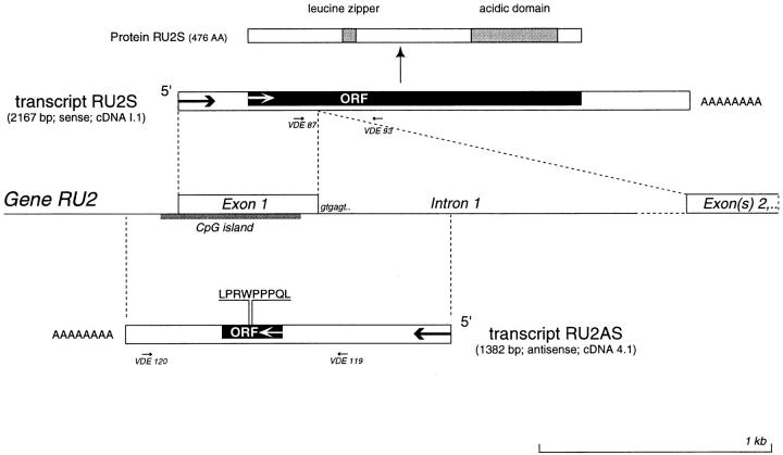
Figure 6.
Partial structure of gene RU2. Black boxes indicate the open reading frames (ORF). The donor splice site of intron 1 is shown. Potentially functional domains of the RU2S protein are indicated. The small arrows indicate the primers used for testing the expression of the two opposite transcripts by RT-PCR: primers VDE87 and VDE93 were used to detect expression of sense transcript RU2S, whereas primers VDE119 and VDE120 were used to detect antisense transcript RU2AS. The 3′ part of intron 1 (dotted line) and the exons that follow it were not sequenced. The genomic sequence of RU2 is available from EMBL/GenBank/DDBJ under accession no. AF181720.

