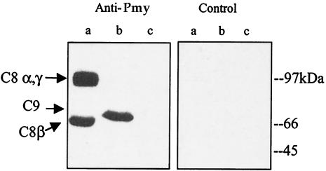
FIG. 3.
Binding of paramyosin to C8 and C9. Purified human C8 and C9 and BSA (1 μg each) were subjected to SDS-8% PAGE and transferred onto a nitrocellulose membrane. After blocking with 5% milk in TBST, the membrane was incubated with recombinant paramyosin (4 μg/ml) in blocking solution for 2 h at 37°C. After two washes with TBST, the membrane was reacted with monoclonal anti-paramyosin antibody (1:4) (Anti-Pmy) or an isotype-matched, unrelated monoclonal antibody (Control) and then incubated with peroxidase-conjugated goat anti-mouse IgG (1:5,000) for 1 h at room temperature. Bands were visualized by enhanced chemiluminescence. Lane a, C8α and C8γ (upper band) and C8β (lower band); lane b, C9; lane c, BSA.

