
Figure 3.
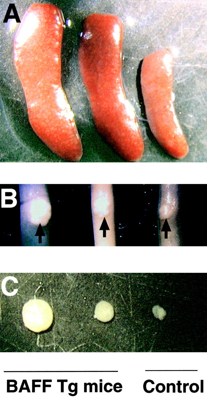
Enlarged spleen, Peyer's patches, and lymph nodes in BAFF-Tg mice. Photograph of (A) spleen, (B) Peyer's patches (indicated with an arrow) on the small intestine, and (C) inguinal lymph nodes of a control mouse (right) and two BAFF-Tg mice (left). Pictures (5×) are representative of at least 12 mice killed for each group.