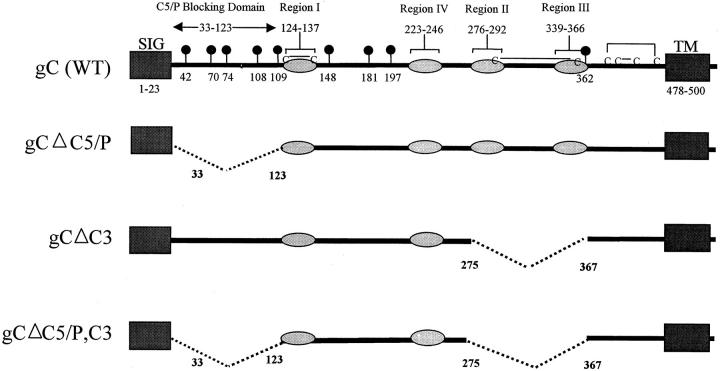
Figure 1.
Stick diagram of wild-type and mutant gC proteins. The top stick figure shows wild-type gC with a signal sequence (SIG), transmembrane domain (TM), the C5/P blocking domain, and four regions (labeled I–IV) shown as shaded ovals that are required for C3 binding. Black balloons show the positions of N-linked glycosylation sites. Cysteines are indicated (C), and lines joining two Cs reflect disulfide bonding pattern. The second stick figure shows the deletion from amino acids 33–123 that eliminates the C5/P blocking domain. The third stick figure shows the deletion from amino acids 275–367 that eliminates C3 binding domain regions II and III. The fourth (bottom) stick figure shows the double deletion that eliminates the C5/P blocking and the C3 binding domain.

