
Figure 5.
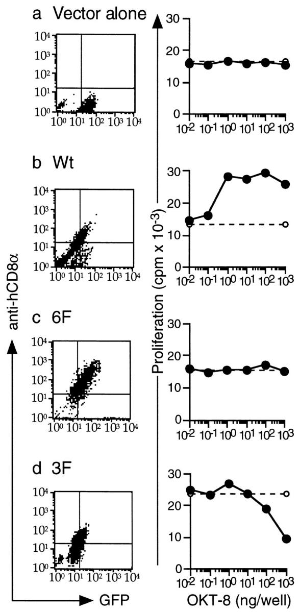
3F can inhibit proliferation in 2.102 TCR-transgenic splenocytes. (a–d) Left panels: expression of Wt, 6F, and 3F in 2.102 T cells. hCD8-ζ mutants Wt, 6F, and 3F were cloned into a bicistronic retrovirus coexpressing GFP. Retrovirus-containing supernatants were produced using the packaging cell line Phoenix E in two independent infections. T cells from the spleens of 2.102 TCR–transgenic, RAG-1−/− mice were harvested, activated in vitro, and infected with retroviral supernatants containing vector alone (a), Wt (b), 6F (c), or 3F (d). T cells were infected on two separate occasions. GFP-positive cells were FACS® sorted, restimulated, and subcloned. Two cell cultures for each construct were then analyzed by FACS® for expression of GFP and hCD8α using RPA-T8–PE (PharMingen). All cell cultures were positive for the expression of GFP, and the Wt-, 6F-, and 3F-transduced T cells also expressed hCD8α. Right panels: 3F can inhibit TCR-mediated proliferation. 3 × 10⁴ 2.102 T cells were stimulated with 5 × 10⁴ CH27 cells prepulsed with Hb(64–76). This led to T cell proliferation as measured with thymidine incorporation after 48 h (dashed lines). Alternatively, increasing doses of OKT-8 were added to the experiment to cross-link the hCD8 mutants (solid lines). This led to an inhibition of T cell proliferation when 2.102 T cells were transduced with 3F (d). OKT-8 had no effect on T cells transduced with empty vector (a) or with 6F (c). The presence of Wt augmented TCR-mediated proliferation (b). The Hb(64–76) doses necessary to obtain intermediate levels of proliferation were determined in previous experiments. Doses were 3 × 10⁻⁶ M for vector alone, Wt, and 3F and 10−6 M for 6F. The data represent the mean of triplicate cultures with SD < 15% of the mean. The experiment was performed three times with similar results.