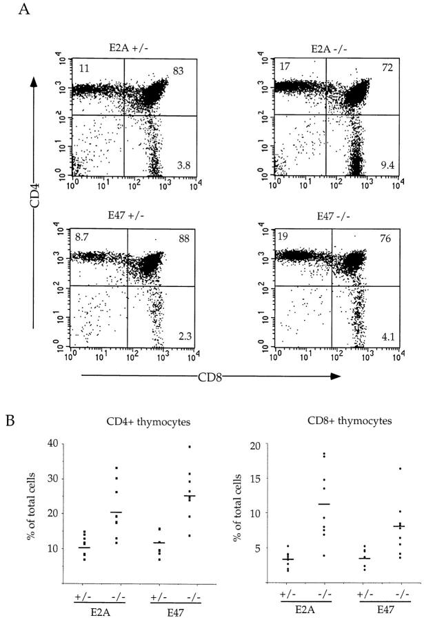
Figure 1.
Increased percentage of SP thymocytes in E2A- and E47-deficient mice. (A) Two-color flow cytometric analysis of thymocytes from 4–6-wk-old E2A- and E47-deficient mice and heterozygous littermates. Thymocytes were analyzed by staining with anti-CD8α and anti-CD4. The numbers in each quadrant indicate the percentage of thymocytes in that population. (B) The percentage of mature CD4+ (left) or CD8+ (right) thymocytes is plotted for eight to nine individual E2A- and E47-deficient mice and heterozygous controls. The horizontal bars indicate the average percentage of CD4+ or CD8+ thymocytes for each genotype.

