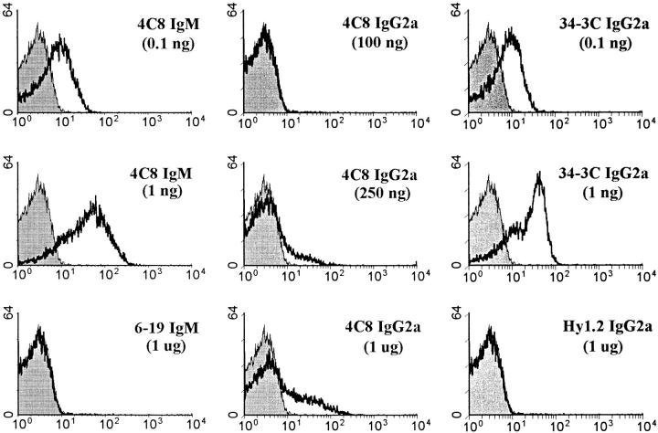
Figure 1.
Flow cytometric analysis of mouse RBCs stained with different amounts of the 4C8 (IgM and IgG2a) and 34-3C (IgG2a) anti–mouse RBC mAbs. RBCs freshly prepared from BALB/c mice were first incubated with different amounts of various mAbs, and then incubated with biotinylated anti–mouse κ chain mAb, followed by PE-conjugated streptavidin. Shaded histograms indicate the background staining with anti–mouse κ chain mAb alone. As controls, the results obtained with isotype-matched control mAbs are shown.

