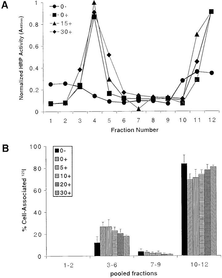
Figure 4.
BCR-bound antigens translocate into rafts after BCR cross-linking. (A) B cells were untreated (0−) or treated with HRP–anti-Ig at 4°C for 1 h, washed, and warmed to 37°C for 0, 15, or 30 min (0+, 15+, and 30+, respectively). The cells were lysed in 1% Triton X-100 in TNEV buffer, the lysates subjected to discontinuous sucrose density gradient centrifugation, and the fractions assayed for HRP activity. (B) CH27 cells were incubated at 4°C for 1 h with 125I–Fab–anti-Ig. During the last 30 min of incubation, the cells were untreated (0−) or treated with anti-Ig to cross-link the BCR and then warmed to 37°C for 0–30 min (0+ to 30+). The cells were lysed in 1% Triton X-100 in TNEV buffer, the lysates were subjected to discontinuous sucrose gradient centrifugation, and the cpm of each fraction was measured and expressed as a percent of the total cell-associated 125I. The average and SEM of three independent experiments is shown.

