
Figure 4.
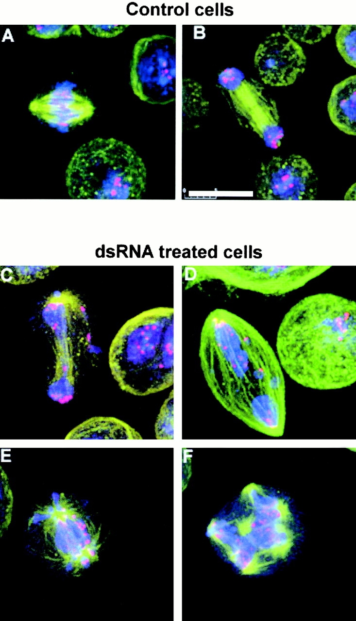
Centromere segregation is defective in Aurora B–depleted cells. (A and B) Control cells at metaphase and anaphase, respectively. (C–F) Abnormal mitoses in aurora B–depleted cells in an anaphase-like state showing centromeric regions (stained red with anti-Prod antibody) that fail to reach the spindle poles. DNA is stained blue and microtubules green. Bar, 10 μm.