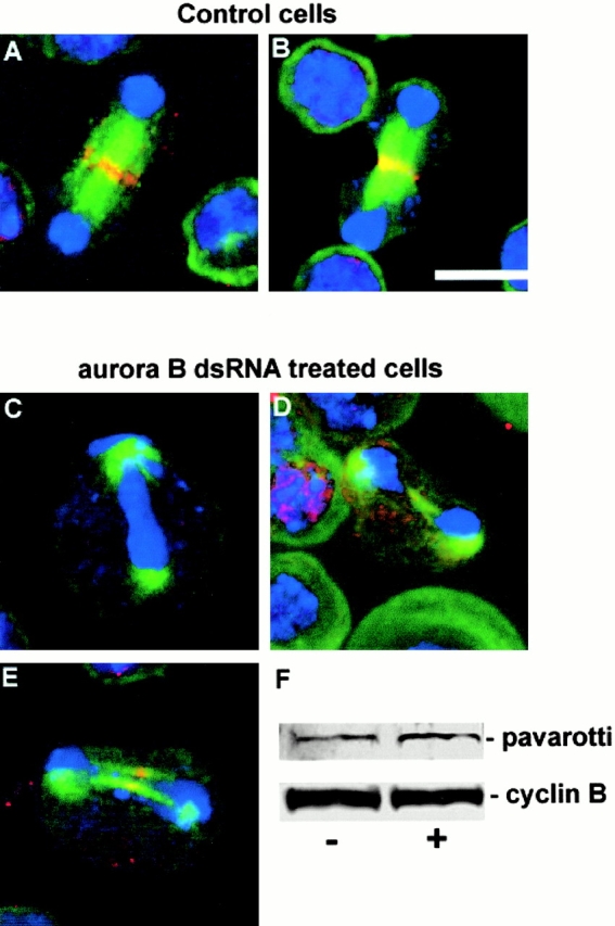
Figure 7.

Failure of Pav-KLP to fully localize to the central spindle in Aurora B–depleted cells. (A and B) Pav-KLP (red) localizes to the central spindle/midbody in control S2 cells at late anaphase (A) and early cytokinesis (B). DNA is blue and microtubules are green. (C–E) Abnormal late mitotic figures in Aurora B–depleted cells in which the density of microtubules in the central spindle region is greatly reduced and in which the Pav-KLP either fails to localize (C) or localizes very poorly (D and E). (F) Western blot showing that levels of Pav-KLP (top) are unchanged from control cells (−) after treatment with aurB dsRNA (+), as are levels of cyclin B (bottom). Bar, 10 μm.
