
Figure 2.
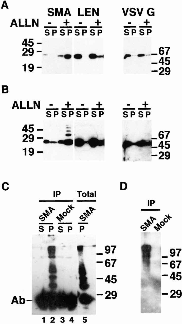
Proteasome inhibitors increase steady state levels of SMA, most of which is found in insoluble aggregates. (A) Transfected cells expressing SMA, LEN, or VSV G proteins were incubated overnight in the absence (−) or presence (+) of 10 μg/ml ALLN. Detergent soluble (S) and pelleted, insoluble (P) fractions were prepared and analyzed by Western blotting with anti–kappa antibody. The SMA fractions were loaded equally; two times more insoluble material was loaded for LEN and VSV G. Insoluble SMA, but not LEN or VSV G, accumulates in the presence of ALLN. (B) Upon long exposure, a ladder of bands is visible in the insoluble fraction, reflecting post-translational modification of SMA. (C) After overnight incubation with 10 μg/ml ALLN, soluble (S) and insoluble (P) fractions from SMA (lanes 1 and 2) or mock-transfected (3 and 4) cells were immunoprecipitated with anti–ubiquitin antibody. Precipitated material was analyzed by Western blotting with anti–kappa antibody. For comparison, total insoluble lysate (lane 5) was also analyzed by Western blotting with anti–kappa antibody. The major, 29-kD band in 1–4 is the light chain from the immunoprecipitating antibody; the 29-kD band in 5 is the monomeric, unmodified form of SMA. (D) After overnight incubation with 10 μg/ml ALLN, insoluble fractions from SMA- or mock-transfected cells were immunoprecipitated with anti–kappa antibody. Precipitated material was analyzed by Western blotting with anti–ubiquitin antibody. Ubiquitinated forms of kappa are detected only in the insoluble fraction from SMA-expressing cells.