
Figure 8.
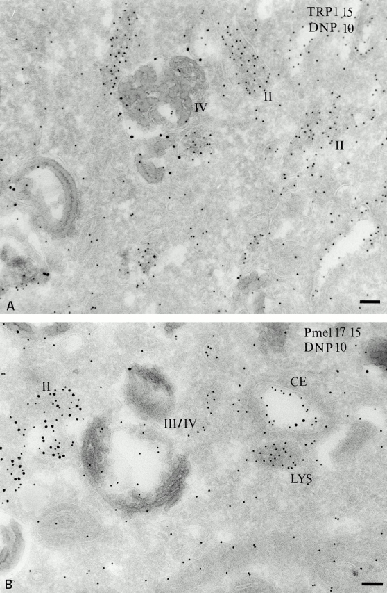
Acidity of melanosomal compartments probed with DAMP. MNT-1 cells were incubated with DAMP for 30 min at 37°C before fixation and processing. (A) DAMP, visualized with anti-DNP antibodies (PAG 10), accumulates primarily in stage II melanosomes displaying characteristic striations. TRP1-positive stage IV melanosomes show less labeling. (B) Double immunogold localization of DAMP (PAG 10) and Pmel17 (PAG 15). DAMP is distributed over the coated endosome, stage II and IV melanosomes, and lysosomes. Bars, 100 nm.