Figure 2.

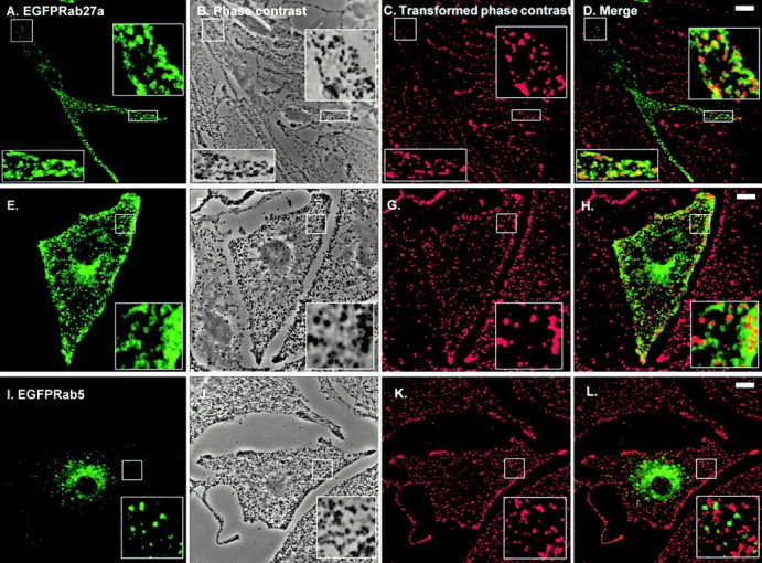
Transiently overexpressed EGFP-Rab27a is associated with pigment granules in melan-a cells. Melan-a cells were transfected with pEGFP-Rab27a (A–H) or pEGFP-Rab5 (I–L) and fixed 48 h later, as described in Materials and Methods with the following modification. After transfection, cells were permeabilized for 5 min before fixation in solution 4 (80 mM K-Pipes, pH 6.8, 5 mM EGTA, 1 mM MgCl2, 0.05% saponin) in order to remove nonmembrane-associated fusion protein. A, E, and I show the distribution of the indicated overexpressed EGFP-Rab fusion protein in melan-a cells. B, F, and J are phase–contrast images of melan-a cells showing the distribution of pigment granules in transfected cells. Phase–contrast images (B, F, and J) of melan-a cells were transformed into pseudocolor (red) images, shown in C, G, and K, using Adobe Photoshop® 4.0 and then merged with the green fluorescence signal emitted by overexpressed EGFP-Rab27a or EGFP-Rab5 proteins, respectively (D, H, and L). Bars, 10 μm.
