
Figure 6.
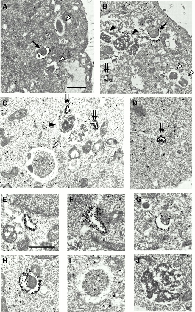
GFP-Apg5 is present on isolation membranes. GFP24 (A–C, E–J) or GKR-1 (D) cells were cultured in Hanks' solution for 2 h and fixed. The localization of GFP-Apg5 was examined by silver-enhanced immunogold electron microscopy using an anti–GFP antibody. In B, an isolation membrane is enclosing a mitochondrion. The isolation membranes (arrows), autophagosomes (open arrowheads) and autolysosomes (closed arrowheads) are indicated. Double arrows indicate small membrane compartments to which GFP-Apg5 extensively localizes. The typical images of these structures are shown at higher magnification (E–J). Bars, 1 μm.