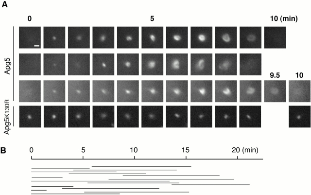
Figure 7.
Formation of autophagosomes is traced with GFP-Apg5. (A) Sequential frames (1-min intervals) of three newly generated GFP-Apg5 spots, or a GFP-Apg5K130R spot. GFP24 or GKR-1 cells were cultured in Hanks' solution for 1 h and directly observed by time-lapse video microscopy. Bars, 1 μm. See supplemental videos of GFP-Apg5 (videos 1–3) and GFP-Apg5K130R (video 4). (B) Duration of each spot of GFP-Apg5 staining was measured for 15 cases that showed circularization in the same field.

