
Figure 8.
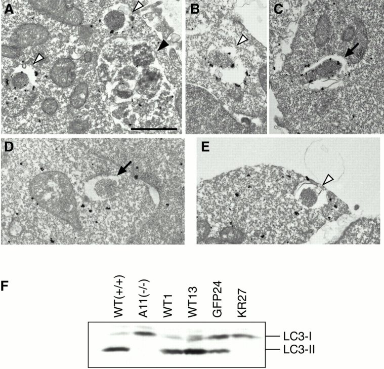
LC3 does not target to membrane in APG5−/− cells. Wild-type ES cells (A–C) and APG5−/− cells (D and E) were cultured in Hanks' solution for 2 h and fixed. Localization of LC3 was examined by silver-enhanced immunogold electron microscopy using an antibody against recombinant LC3. The isolation membrane (arrow), autophagosomes (open arrowheads) and autolysosome (closed arrowhead) are indicated. Bar, 1 μm. (F) Total lysates from APG5 +/+ and APG5 −/− cells and their various stable transformants cultured in Hanks' solution for 2 h were subjected to immunoblot analysis with anti–LC3 antibody. Positions of LC3-I and LC3-II are indicated.