
Figure 2.
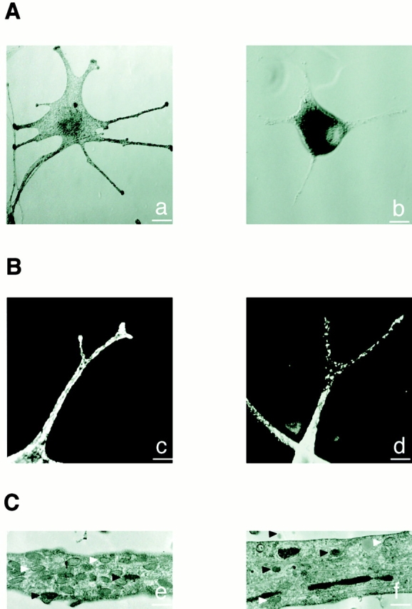
Melanosome distribution is markedly impaired in GS melanocytes. (A) Phase–contrast microscopy images of a normal (a) and GS (b) melanocyte. (B) Confocal immunofluorescence microscopy images of a normal (c) and GS (d) melanocyte labeled for melanosomes with an anti–TRP-1 antibody. (C) Electron microscopy images of a dendritic extension of a normal (e) and GS (f) melanocyte. Note that despite the reduction of melanosome number, both partially (white arrowheads) and fully pigmented (black arrowheads) melanosomes are found in the dendrite of the GS melanocyte. Bars: (A) 25 μm; (B) 5 μm; (C) 1 μm.