
Figure 3.

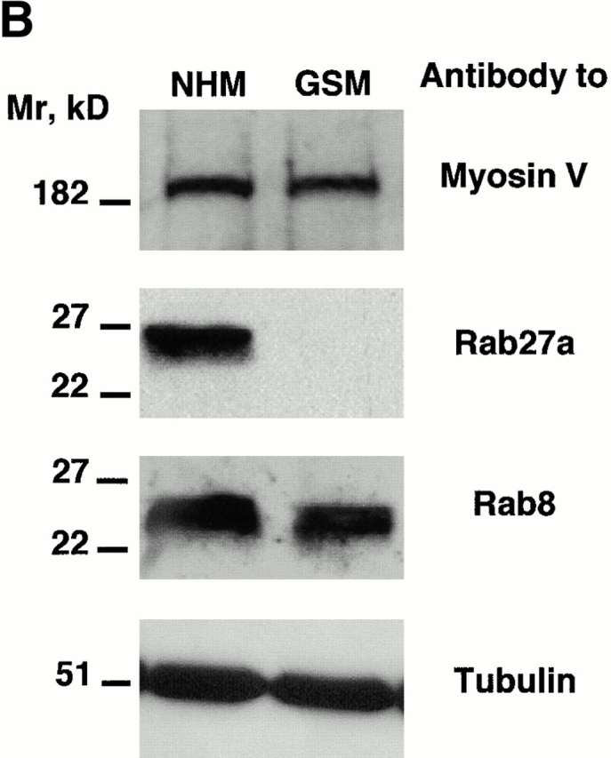
Absence of Rab27a expression in GS melanocytes. (A) Immunofluorescence labeling of myosin-V in normal (a) and GS (b) melanocytes, and of Rab27a in normal (c) and GS (d) melanocytes. For Rab27a labeling, Hoechst staining was used to visualize the nuclei. (B) Western blot analysis. Membranes were blotted with a polyclonal antimyosin-V antibody (top). Membranes were blotted first with a monoclonal anti-Rab27a antibody (middle). After stripping, membranes were reblotted with a monoclonal anti-Rab8 antibody. Membranes were blotted with a monoclonal antitubulin antibody to control the equal amount of protein in each lane (bottom). Bars, 30 μm.