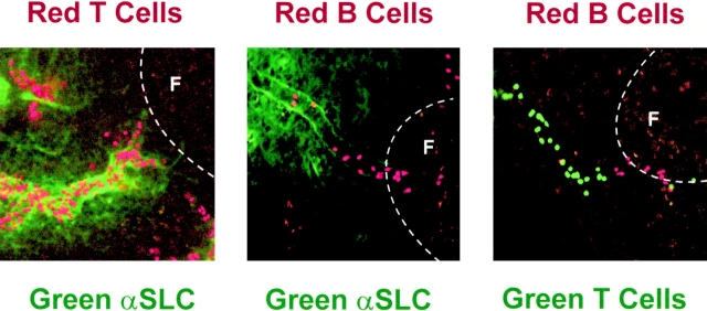
Figure 7.
Localization of lymphocytes in PP-HEVs. Micrographs of accumulated lymphocyte subpopulations in PPs. (Left panel) T cells accumulated in SLC+ interfollicular segments of HEVs. (Middle panel) B cells in SLClow HEV segments associated with primary follicles. (Right panel) Segregation of sites of preferential T and B cell accumulation in relation to follicles. TRITC-labeled T (left) or B (middle and right panels) cells and CMFDA-labeled T cells (right panel) were observed in HEVs of normal animals as in the legend to Fig. 4. In the left and middle panels, after lymphocyte accumulation for ∼10 min (see Materials and Methods), 60 μg Alexa™ 488–conjugated anti-SLC mAb was injected intravenously to illuminate SLC-expressing HEV segments. Animals were killed, and whole PPs were removed for examination by confocal microscopy. The location of follicles is indicated (F) (20× objective).

