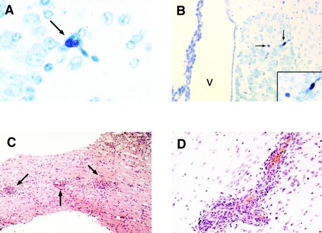
Figure 2.
Histologic analyses of CNS tissues in WBB6/F1 +/+ mice. After sacrifice of the animals, brains, spinal columns, and other organs were removed and preserved in 10% neutral buffered formalin. Paraffin-embedded tissue sections were stained with Giemsa (A and B) or hematoxylin and eosin (C and D). (A) Mast cell (arrow) located within the thalamic border region of the habenula; ×40. (B) Two mast cells (arrows) located in the habenula. The third ventricle is also noted (V); ×20. Inset, the same two mast cells ×40. (C) Multiple inflammatory infiltrates (arrows) found in spinal cord section of a diseased animal; ×10. (D) Focal inflammatory infiltrate found in the brain parenchyma of a diseased animal; ×40.

