
Figure 6.
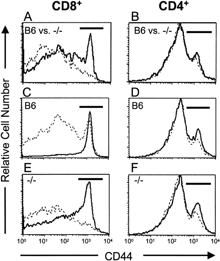
IL-15−/− mice have a reversible defect in CD8+ memory phenotype (CD44hi) T cells. Splenocytes and LN cells were incubated with mAb specific for CD4, CD8, and CD44. Gated populations of CD8+ or CD4+ cells were analyzed for expression of CD44. The CD44hi populations are indicated by the horizontal bars on each histogram. (A and B) CD44 expression was analyzed on CD8+ and CD4+ splenocytes and LN cells from individual 15-wk-old naive C57BL/6 (B6) or IL-15−/− female mice (n = 4/group). Similar patterns were observed in both LNs (not shown) and spleen. (A) CD44 expression on CD8+ splenocytes from control (solid line) or IL-15−/− (dashed line) mice. The mean percentage ± SEM of CD44hi cells within the CD8+ population was 19 ± 2% for control mice and 6.5 ± 0.5% for IL-15−/− mice. (B) CD44 expression on CD4+splenocytes from control (solid line; 22.2 ± 2% CD44hi) or IL-15−/− (dashed line; 22 ± 1% CD44hi) mice. (C–F) 9-wk-old C57BL/6 or IL-15−/− female mice (n = 3/group) were injected intraperitoneally with PBS (dashed line) or 10 μg human IL-15 (solid line) once daily for 7 d. Spleens and LNs were removed 24 h after the last injection. Single cell suspensions from individual mice were analyzed for CD4, CD8, and CD44 expression as described above. Similar changes were observed in both LNs (not shown) and spleen (see Table for summary of data). (C and E) CD44 expression on CD8+ splenocytes from PBS- or IL-15–treated control (C) or IL-15−/− (E) mice. (D and F) CD44 expression on CD4+ splenocytes from PBS- or IL-15–treated control (D) or IL-15−/− (F) mice.