Figure 1.

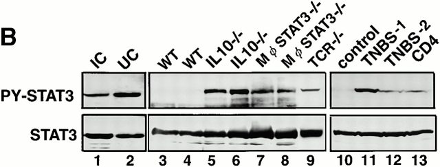
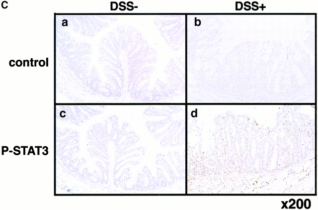
Expression and activation of STATs in human and mouse colon tissues. (A) Colon tissue extracts from the normal region (NR) of colon cancer patients (lane 1), those from UC or CD patients (lanes 2–4), or those from Balb/c mice treated without (day 0) or with 4% DSS for 7 d (lanes 5 and 6) were immunoblotted with antiphospho-STATs (PY-STAT) or anti-STAT antibodies. As a positive control (pos), IFN-γ–treated NIH-3T3 cells (for STAT1), LIF-treated CMT cells (STAT3), IL-3–treated Ba/F3 cells (STAT5), and IL-4–treated 32D cells (STAT6) are shown in lane 7. We analyzed more than seven patients and three DSS-treated mice, obtaining similar results. (B) STAT3 activation in UC and IC patients (lanes 1 and 2) as well as several mouse models (lanes 3–13). Colon samples were obtained from 10–15-wk-old WT C57BL6 mice (lanes 3 and 4), 16-wk-old IL-10−/− mice (lanes 5 and 6), 9-wk-old Mφ-STAT3−/− mice (lanes 7 and 8), 15-wk-old TCR−/− mouse (lane 9), TNBS-treated (lanes 11 and 12) or untreated (lane 10) mice, and CD4+ T cell–transplanted SCID mouse (lane 13). (C) Immunohistochemical detection of activated STAT3 in DSS-induced colitis. C57BL6 mice were treated with (b and d) or without (a and c) 2% DSS for 5 d, and colon tissues were fixed with 10% formalin. Slides with sectioned tissues of 7-μm thickness were immunostained with anti–phospho-STAT3–specific antibodies (c and d) or control IgG (a and b) and were lightly stained with hematoxylin.


