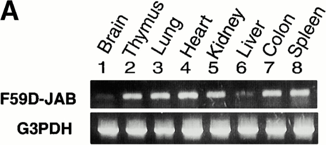
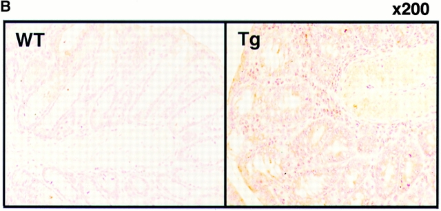
Figure 7.
Expression of F59D-JAB in Tg mice. (A) RT-PCR analysis for tissues from F59D-JAB Tg mouse. 1 μg total RNA from each tissue was used as a template to detect F59D-JAB and control G3PDH mRNAs. Similar levels of expression were observed in the other three Tg lines. (B) Immunohistochemical detection of F59D-JAB in the colon of WT littermate or F59D-JAB Tg mouse (Tg).


