Figure 4.
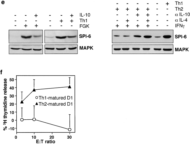
Th1 induces, while Th2 and IL-10 block SPI-6 induction in D1. (a) 5 × 10⁴ OVA-specific Th1 and Th2 were incubated with 106 D1 cells for 48 h. Maturation of OVA peptide–loaded D1 cells was measured by B7-2 expression. (b–d) 106 D1 cells were incubated with 5 × 10⁴ Th1 or Th2 cells, peptide, anti-CD40L, and anti–IL-10 as indicated and tested for IL-12p40 secretion by ELISA and SPI-6 expression by Western blotting after 24 h. Lysates from equivalent numbers of cells were applied on SDS-PAGE (0.5 × 10⁶ cells). Equal protein loading was confirmed by ponceau S staining as well as by a control Western for MAPK (bottom). Please note that D1 cells were incubated with Th cells at a ratio of 20:1, resulting in a 20-fold lower protein content in the samples that contained Th cells only (right lanes). (e) Anti-CD40 (FGK) or Th1-induced SPI-6 expression in D1 cells was determined after 48 h in the absence or presence of IL-10 (left). The right panel represents SPI-6 expression in D1 cells after incubation with Th2 cells in the presence or absence of IFN-γ, anti–IL-10, and anti–IL-4 as indicated. As a control the Th1-induced SPI-6 expression is shown on the far right. (f) DNA fragmentation of Th1- (circles) or Th2 (triangles)-matured D1 cells (E1B-peptide loaded) was tested with the E1B-specific CTL clone for 6 h as described in the legend to Fig. 1 a.


