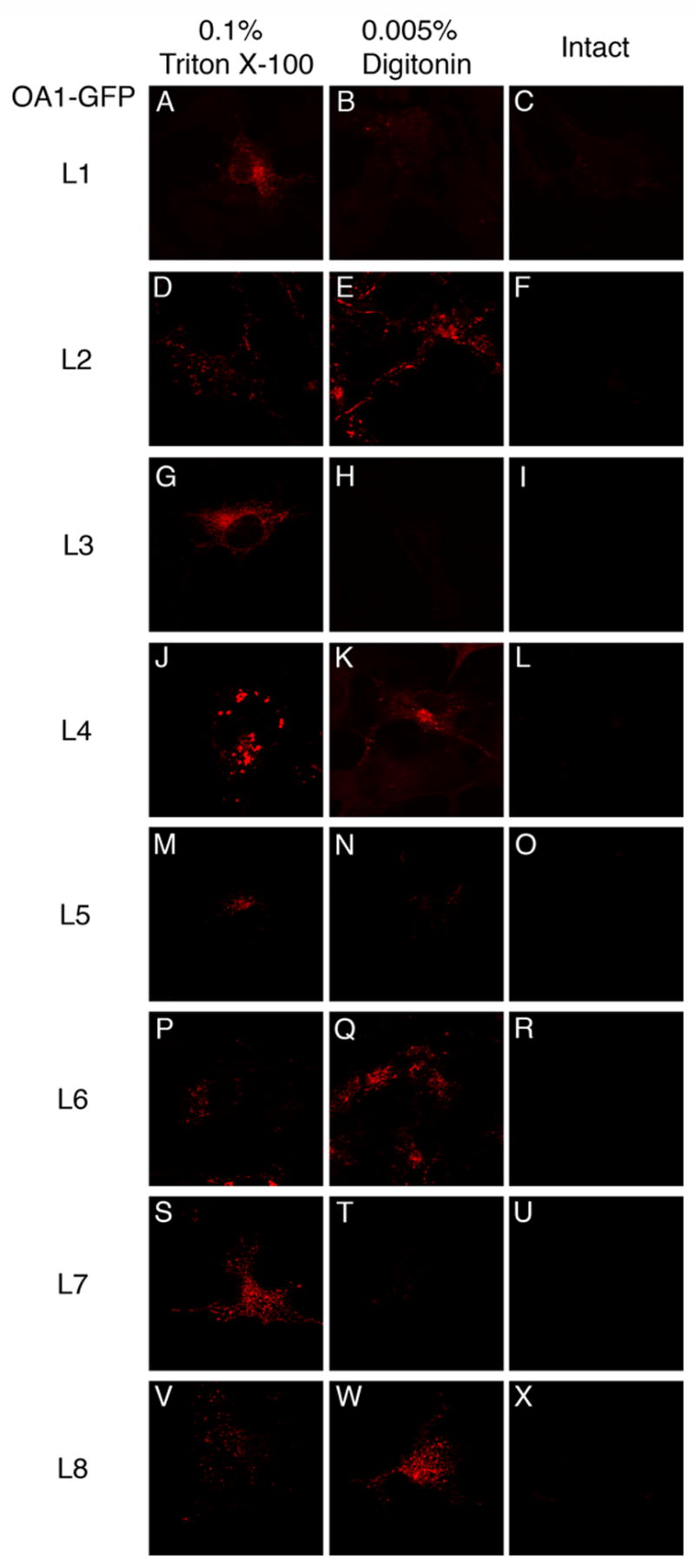
Fig. 5. Orientation of HA-tag inserted in OA1-GFP derivatives as assessed by immunofluorescence of partially permeabilized cells.

COS-1 cells transiently expressing OA1-GFP derivatives were fixed with formaldehyde and partially permeabilized with the condition as in Fig. 4. OA1-GFP derivatives expressed in COS-1 cells were: OA1-GFP (L1) (A, B, and C); OA1-GFP (L2) (D, E, and F); OA1-GFP (L3) (G, H, and I) ; OA1-GFP (L4) (J, K, and L); OA1-GFP (L5) (M, N, and O); OA1-GFP (L6) (P, Q, and R); OA1-GFP (L7) (S, T, and U); OA1-GFP (L8) (V, W, and X). HA-tag inserted to L2, L4, L6, or L8 (E, K, Q, W) was labeled by anti-HA, while that to L1, L3, L5, or L7 (B, H, N, T) was not.
