
Figure 2.
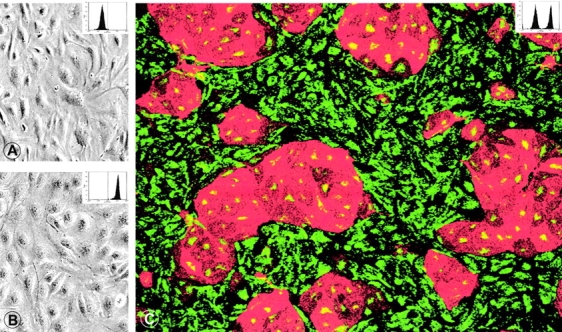
BECs and LECs cannot be distinguished by conventional light microscopy when grown separate (A, BECs; B, LECs). (C) In mixed cultures, immunolabeling with anti-podoplanin (TRITC, red) and anti-vWF (FITC, green) reveal multicellular islands of podoplanin+/vWF+ LECs surrounded by podoplanin−/vWF+ BECs. BECs express more vWF than LECs (see also Fig. 3 A). Original magnification in A and B: ×300; C: 250. To confirm their phenotypic stability, cultured LECs and BECs were harvested, labeled with anti-podoplanin IgG, and analyzed by FACS ® (insets in A–C; dotted line denotes the cut-off for podoplanin positivity). Cultured BECs (A) were homogeneously podoplanin-negative, cultured LECs (B) exclusively podoplanin-positive, while the mixed EC population (C) contained both qualities.