Figure 2.
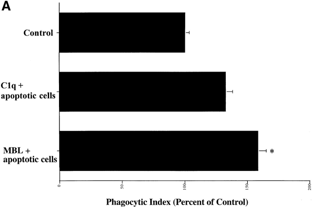
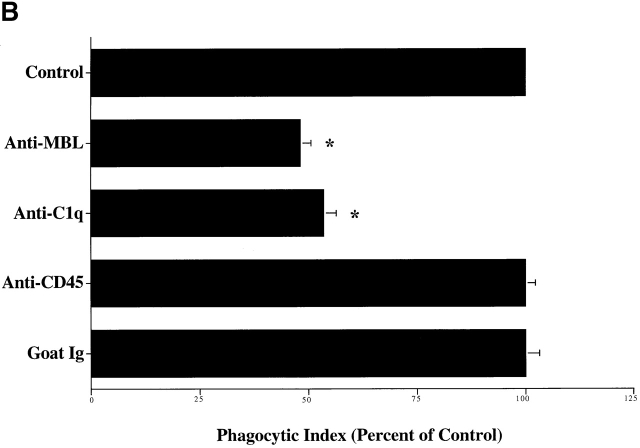
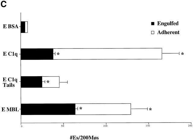
C1q and MBL facilitate particle clearance. (A) Pretreatment of apoptotic cells with C1q or MBL: apoptotic cells (5 × 10⁶) were incubated with 50 μg of purified C1q or MBL. These cells were tumbled for 30 min at 37°C and washed with HBSS before addition to macrophages for a phagocytosis assay (as described in Materials and Methods). n = 4 ± SEM (control mean phagocytic index = 44.2 ± 6.6, P < 0.05). (B) Antibody against human C1q and against human MBL inhibits uptake of apoptotic cells into HMDMs. HMDMs were treated with 10 μg antibody (polyclonal goat anti–human C1q, goat control antibody, mouse monoclonal anti-CD45 antibody as control, and monoclonal mouse anti–human MBL, shown) for 30 min at 37°C. Media was then aspirated, and apoptotic cells were added for 1 h for a phagocytosis assay. n = 4 ± SEM (control mean phagocytic index = 37.0 ± 6.1, *P < 0.005). (C) C1q, C1q Tails, or MBL Ebab are ingested by HMDMs. Single ligand coated particles coated with control protein, MBL, C1q, or the collagenous tail fragment of C1q were fed to HMDMs for 20 min at 37°C. The cells were then washed, fixed, and counted (as described in the Materials and Methods section). n = 3 ± SEM. P < 0.005 Engulfed; P < 0.003 Adherent.


