Figure 4.
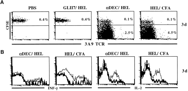
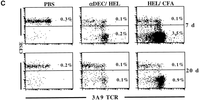
CD4+ T cells divide in response to antigen presented by DCs in vivo, produce IL-2 but not IFN-γ, and are then rapidly deleted. (A) CFSE labeled CD45.1+ 3A9 T cells were transferred into B10.BR and 24 h later, the recipients were injected subcutaneously in the footpads with αDEC/HEL (0.2 μg), GL117/HEL (0.2 μg), HEL peptide in CFA, or PBS. CD4+ T cells were purified by negative selection from regional LNs 3 d after challenge with antigen and analyzed by flow cytometry. The plots show staining with 1G12 anti-3A9 and CFSE intensity on gated populations of CD4+CD45.1+ cells. The numbers indicate the percentage of CFSE high (undivided) and CFSE low (divided) CD4+ T cells. The results are from one of two similar experiments. (B) T cells produce IL-2 but not IFN-γ in response to antigens presented on DCs under physiological conditions. 3A9 cells were transferred into B10.BR mice and 24 h later the recipients were injected subcutaneously in the footpads with αDEC/HEL (0.2 μg), GL117/HEL (0.2 μg), HEL peptide in CFA. CD4+. Histograms show staining with anti–IL-2 and anti–IFN-γ on gated populations of 3A9+CD4+ cells. The thick lines indicate PBS control. (C) Same as in panel A but analysis performed 7 or 20 d after antigen administration.


