
Figure 3.
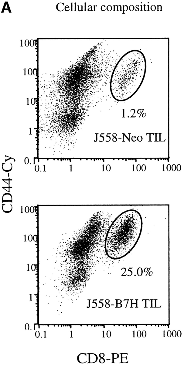
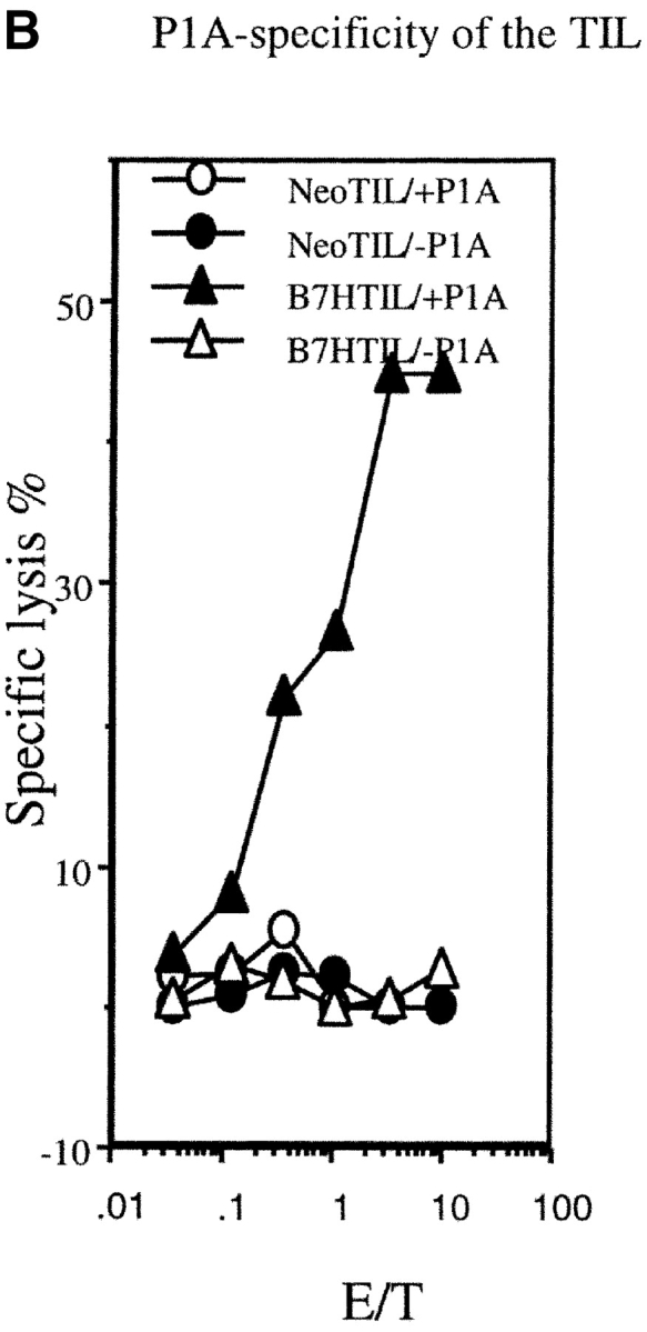

B7H increases P1A-specific CTL in the TIL from wild-type BALB/c mice. The TIL were enriched by depletion tumor cells as described (reference 12). The cytotoxicity of the freshly isolated TIL were determined using either P1A-pulsed or unpulsed P388D1, or the J558-Neo and J558-B7H cells as targets. (A) Substantial increase of activated CD8 T cells among the TIL. The TIL were stained with PE-conjugated anti-CD8 and Cy-conjugated anti-CD44 mAbs and analyzed by flow cytometry. (B) P1A-specific CTL in the TIL from the J558-B7H, but not the J558-Neo tumors. Freshly isolated TIL were used as effectors while the P1A-pulsed and unpulsed P388D1 were used as targets. (C) TIL from J558-B7H (right panel), but not those from J558-Neo (left panel), lysed both J558-B7H and J558-Neo tumors. As in B, except that the J558-Neo and J558-B7H were also used as targets. Data shown are representative of at least three independent experiments. TIL used in A and B were isolated at day 24 of tumor injection, while those used in C were isolated on day 20 of tumor cell injection.