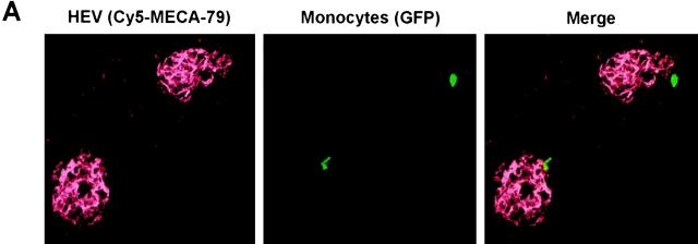
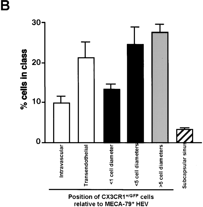
Figure 3.
Monocytes home to inflamed PLNs via HEVs. Adoptive transfer of CX3CR1+/GFP PBMCs into wild-type recipients was performed 5 d after induction of skin inflammation as described in Fig. 2. Draining PLNs were removed after 1 h and prepared for confocal microscopy using appropriate filters for detection of GFP+ monocytes (green) and HEVs, which were visualized using anti-PNAd mAb MECA-79 and Cy5-labeled anti–rat IgM (red). (A) Representative confocal micrographs of homed GFP+ monocytes in close proximity to MECA-79+ HEVs. (B) The frequency distribution of homed GFP+ monocytes relative to MECA-79+ HEVs and the subcapsular sinus was determined. Data are expressed as mean ± SEM of the percentage of GFP+ cells in each class. n = 90 random sections of five inflamed PLNs from two independent experiments.


