Figure 2.

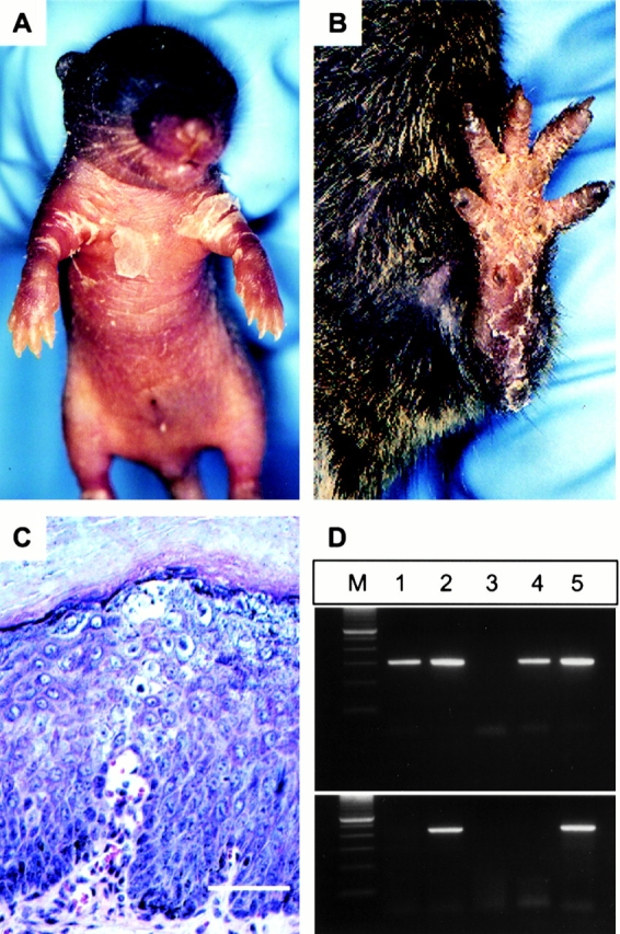
Inducible mouse model for EHK. (A) Bigenic +/mutneo. CrePR1 pup after four topical applications of RU486. Scaling around the arm folds after rupture of the blister. No blisters developed in untreated bigenic +/mutneo.CrePR1 pups or +/mutneo or CrePR1 pups treated with RU486 (data not shown). (B) Same mouse 3 mo after induction. Note the thick brownish hyperkeratoses on the paws. (C) Light micrograph of lesional skin from B. Note the thickened stratum corneum, vacuolization, and translucency in the suprabasal layer. Hematoxylin and eosin, bar = 50 μm. (D) PCR analysis of keratinocytes captured by LCM. PCR analysis to detect the loxP site (top panel) and neo cassette (bottom panel). 1, suprabasal cells from previously treated area; 2, untreated area; 3, wild-type control mouse; 4, +/mutloxP mouse; and 5, +/mutneo mouse. M, DNA size marker.
