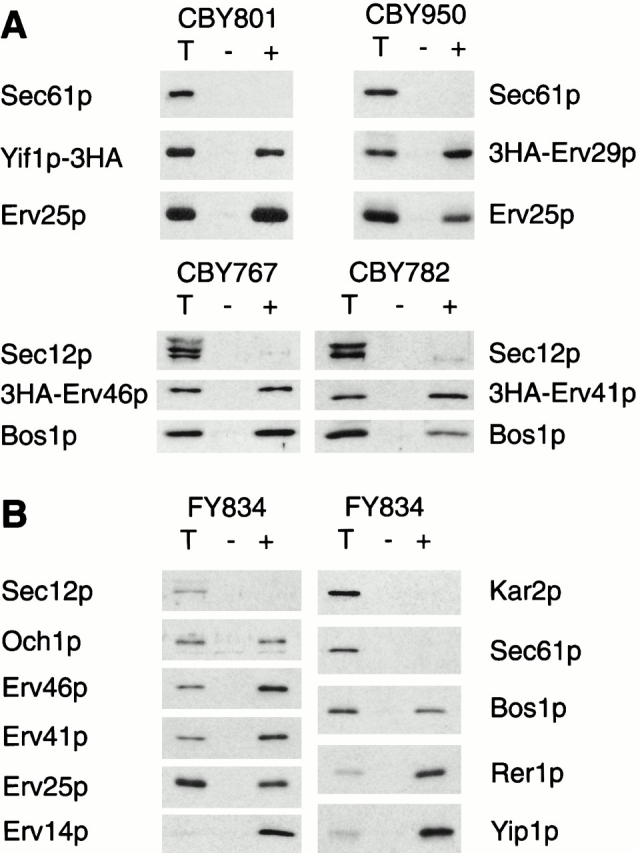
Figure 2.

Selective packaging of Erv proteins into COPII-coated vesicles in vitro. (A) In vitro budding reactions with microsomes prepared from strains expressing tagged versions of Erv proteins: Yif1p-3HA (CBY801), 3HA-Erv29p (CBY950), 3HA-Erv46p (CBY767), and 3HA-Erv41p (CBY782). One tenth of a total reaction (T), budded vesicles isolated after incubation with COPII proteins (+), or a mock reaction without COPII proteins (–) were separated on a 12.5% polyacrylamide gel. Tagged proteins were visualized by immunoblot with an anti-HA antibody, and Sec61p or Sec12p (ER resident proteins) as negative controls and Erv25p (Erv protein) or Bos1p (v-SNARE) as positive controls were detected using polyclonal antisera. (B) In vitro budding reactions with FY834 wild-type microsomes. The same budding protocol as in A was used. Proteins were detected with polyclonal antisera against Sec12p, Kar2p, and Sec61p (ER residents), Bos1p (v-SNARE), Och1p (early Golgi marker), and the Erv proteins Erv46p, Erv41p, Erv25p, Erv14p, Rer1p, and Yip1p.
