
Figure 6.
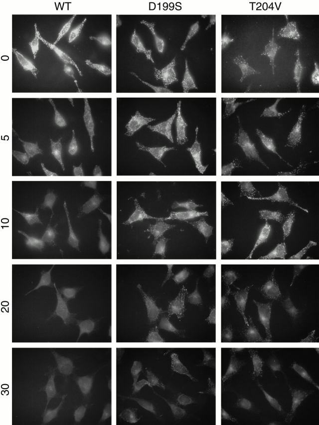
Internalized Tfn exhibits prolonged trafficking through punctate structures in hsc70D199S- and hsc70T204V-expressing cells as visualized by epifluorescent microscopy. A 5-min pulse of Alexa488-Tfn (0 min) was chased with unlabeled Tfn and followed for 5, 10, 20, and 30 min. Cells were fixed and processed for fluorescence microscopy as described in Materials and Methods. All images were collected with 2-s exposures. WT, wild type.