
Figure 8.
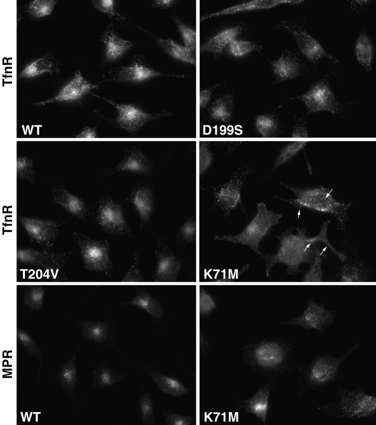
Steady state distribution of TfnR and MPR in wild-type and mutant hsc70–expressing cells. The cellular localization of PM- and TGN-cycled receptors were visualized by epifluorescent microscopy. Anti–TfnR D65 and anti-MPR antibodies were used to label adenovirally infected cells after their fixation. Arrows indicate the tubular TfnR-labeled structures observed in hsc70K71M-expressing cells. WT, wild type.