Figure 4.
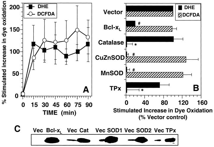
Anti-CD3–induced ROS generation in human T blasts. (A). Kinetics of anti-CD3–stimulated DCFDA/DHE oxidation human T blasts. Human T blasts were stimulated with anti-CD3 as described in Materials and Methods and the oxidation of DCFDA (○) and DHE (▪) was determined by FACS® analysis. The data are expressed as the percent stimulated increase in mean channel fluorescence of DCFDA/DHE over unstimulated controls and represent the average of at least five separate experiments (± SEM). (B) Effect of protein overexpression on DCFDA/DHE oxidation in human T blasts. Human T blasts were transfected with empty vector (Vector) or expression vectors encoding BCl-xL, catalase, CuZnSOD, MnSOD, or TPx at a 2:1 ratio with one encoding GFP or RFP as a transfection marker. After 16 h incubation, anti-CD3–induced DCFDA (hatched bars) or DHE (closed bars) oxidation was measured as described in Materials and Methods and the legend to Fig. 3. DHE oxidation was measured in GFP+ cells while DCFDA oxidation was measured in RFP-transfected cells, both after 15 min anti-CD3 stimulation and was normalized to Vector control. The data represent the average of at least four separate experiments (± SEM). *Significantly different from that in Vector transfected samples (P < 0.05). #Significantly different from that in Vector transfected samples (P < 0.05). (C) Western blot confirmation of protein overexpression. Human T blasts were transiently transfected with the indicated mammalian expression vectors in twofold excess over a marker plasmid expressing a chimeric CD8-Igγ2a protein. After 16 h incubation, viable CD8+ cells were purified by magnetic bead selection. Protein overexpression was determined in positively selected cells by Western blot as described in Materials and Methods.

