
Figure 1.
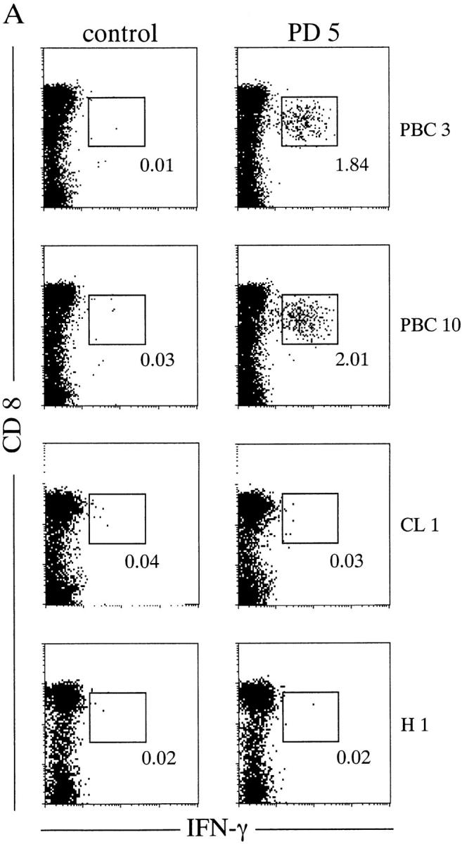

PD5-specific CD8+ T cells from in vitro–cultured PBMCs. PBMCs from HLA-A2+ donors were cocultured with PD5-loaded APCs (autologous immature DCs) for 12 d, then restimulated with PD5 or a control peptide in the presence of brefeldin A, followed by intracellular staining for IFN-γ. (A) IFN-γ staining of samples from two PBC patients, PBC3 and PBC10, and two control donors, CL1 (alcoholic liver disease) and H1 (a healthy donor). Displayed in the dot plots are cells gated for lymphocyte population by forward-scattering and side-scattering and the CD4− population. The cells within the box are considered IFN-γ+. The number next to the box is the percentage of IFN-γ+ cells in the CD8+ T cell population. (B) Frequency of PD5-specific CD8+ T cells in PD5-stimulated PBMC cultures derived from all 20 donors. The cut off value for a positive response was determined as 0.150%, or 3 SD above the mean percentage of IFN-γ producing cells in all 20 samples restimulated with the control peptide. A significant number of IFN-γ–producing cells were detected after restimulation with PD5 in 10/12 (83%) PBC patients but in 0/8 control individuals (P < 0.0007).