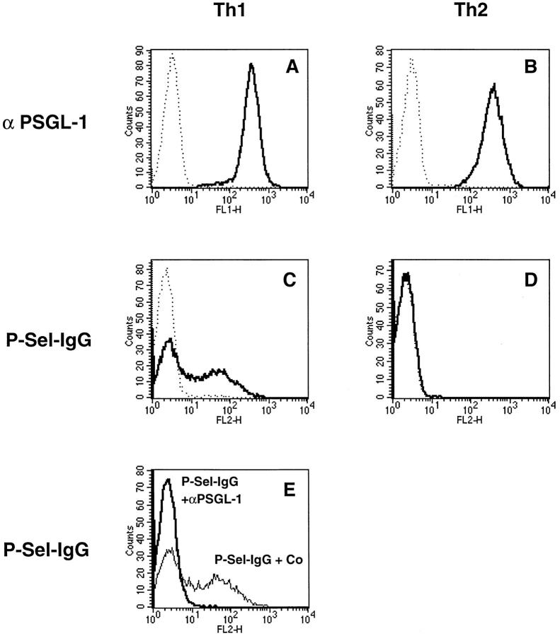
Figure 2.
FACS® analysis of Th1 and Th2 cells with P-selectin–Ig and antibodies against PSGL-1. Th1 and Th2 cells (as indicated) were analyzed by flow cytometry either with affinitypurified rabbit antibodies against mouse PSGL-1 (A and B, solid lines) or with P-selectin–Ig (C and D, solid lines). Dotted lines show negative control staining either with nonimmune rabbit IgG (A and B) or with human IgG (C and D). E shows the staining of Th1 cells with P-selectin–IgG after preincubation of the cells either with nonimmune rabbit IgG (faint line) or with affinitypurified rabbit anti–PSGL-1 antibodies (bold line). P-selectin–Ig was detected with PE-conjugated F(ab′)2 donkey anti–human IgG, and rabbit antibodies were detected with FITC-conjugated goat anti–rabbit IgG.

