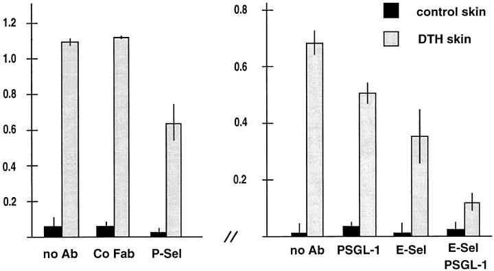
Figure 4.
Partial inhibition of Th1 cell immigration into inflamed skin by antibodies against PSGL-1. Radiolabeled Th1 cells were injected together with PBS (no Ab) or the same buffer containing 100 μg of nonimmune rabbit IgG Fab fragments (Co Fab), 100 μg of affinity-purified anti–PSGL-1 Fab fragments (PSGL-1), 200 μg of mAb RB40 against mouse P-selectin (P-Sel), 200 μg of mAb UZ4 against mouse E-selectin (E-Sel). Immigration of cells into the noninflamed control skin region of the same mice is depicted as solid bars. For each determination, four mice were analyzed. Experiments shown by the left graph were performed with a different preparation of Th1 cells than the experiments depicted by the right graph. Numbers on the left refer to the percentage of injected cells that were found in the analyzed skin area of 2.5 cm2.

