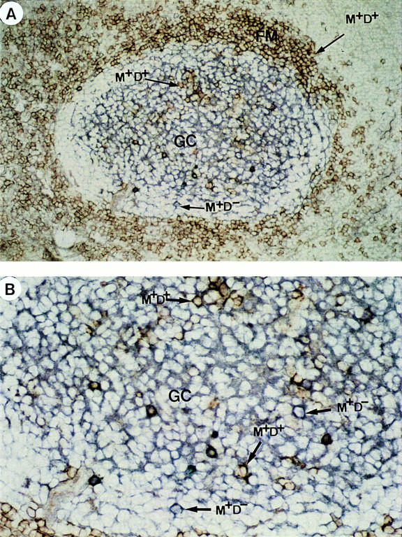
Figure 4.

Identification of IgM+IgD+ and IgM+IgD− B cells within GC: Double anti– IgD (red) and anti–IgM (blue) staining of a tonsil section. (A) shows a secondary lymphoid follicle that contains purple IgD+IgM+ follicular mantle B cells. Within the GC, IgM immune complexes (blue) on follicular dendritic cells can be observed. In addition, scattered purple IgM+IgD+ B cells and blue IgM+IgD− B cells can be found (×100). (B) Shows a part of this GC at ×200 magnification.
