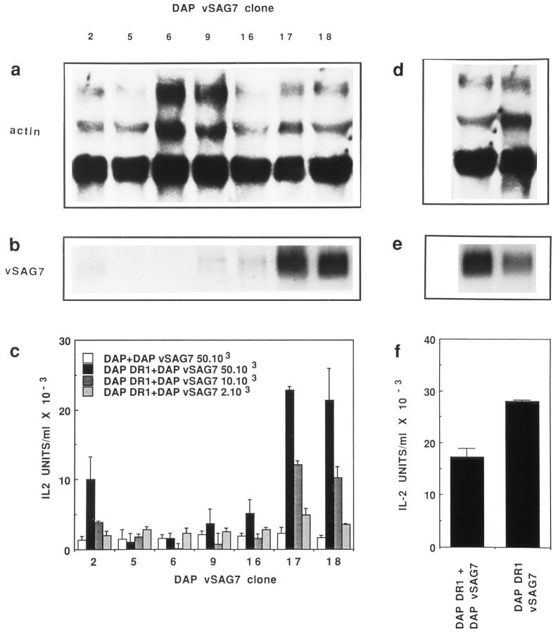
Figure 1.
In vitro transfer of vSAG7 from class II− cells to class II+ cells. (a and b) Expression levels of vSAG7 and actin mRNA in different clones of DAP cells transfected with vSAG7. RNA was extracted from cloned DAP vSAG7 transfectants. 10 μg of RNA were run on a 1.2% agarose-formaldehyde gel and transferred on a nylon membrane. Membranes were then hybridized with either actin (a) or vSAG7 (b) radiolabeled probes and exposed for 24 h. Each track represents an individual clone. (c) Murine fibroblasts transfected with vSAG7 can stimulate T cell hybridomas in the presence of DAP DR1 cells. 6 × 10⁴ DAP DR1 cells and 6 × 10⁴ Kmls 13.11 cells were incubated overnight with either 5 × 10⁴ DAP cells or various amounts of DAP vSAG7 cells. Supernatants were then harvested and tested for IL-2 activity. (d and e) Levels of vSAG7 expression by DAP vSAG7 clone 18 and DAP DR1 vSAG7 are comparable. RNA was extracted from DAP vSAG7 clone 18 and DAP DR1 vSAG7 clone 3B2, run on a 1.2% agarose-formaldehyde gel and hybridized with actin (d) or vSAG7 ( f ) radiolabeled probes. ( f ) Levels of stimulation in the transfer assay (DAP vSAG7 + DAP DR1) and in direct presentation (DAP DR1 vSAG7) are comparable. 6 × 10⁴ Kmls 13.11 cells were cocultured in 250 μl either with 6 × 10⁴ DAP DR1 cells and 5 × 10⁴ DAP vSAG7 clone 18 cells, or with 6 × 10⁴ DAP DR1 vSAG7 cells. Supernatants were then harvested and tested for IL-2 production.

