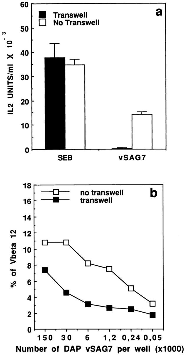
Figure 6.

(a) Transfer of vSAG7 requires cell proximity to stimulate murine T cell hybridoma. 5 × 10⁴ DAP vSAG7 cells or 75 pg SEB were disposed in the upper chamber while 6 × 10⁴ DAP DR1 cells and 6 × 10⁴ Kmls 13.11 cells were disposed in the bottom chamber, separated by a 0.2-μm porous membrane. As a control, 5 × 10⁴ DAP vSAG7 cells or SEB were disposed with the other cells in absence of compartmentalization. Assays were then performed as previously described. (b) The transferred fragment of vSAG7 can cross the membrane and stimulate human PBMCs. Different concentrations of DAP vSAG7 were disposed in the upper chamber while 106 human PBMCs were disposed in the bottom chamber. Percentage of CD4+ blasts expressing Vβ12 or Vβ6.7 was measured by flow cytometry after 7 d of culture. As a control, DAP vSAG7 cells and human PBMCs were disposed in absence of compartmentalization. Results are illustrated as dot plots with Vβ in x axis and CD4 in y axis.
