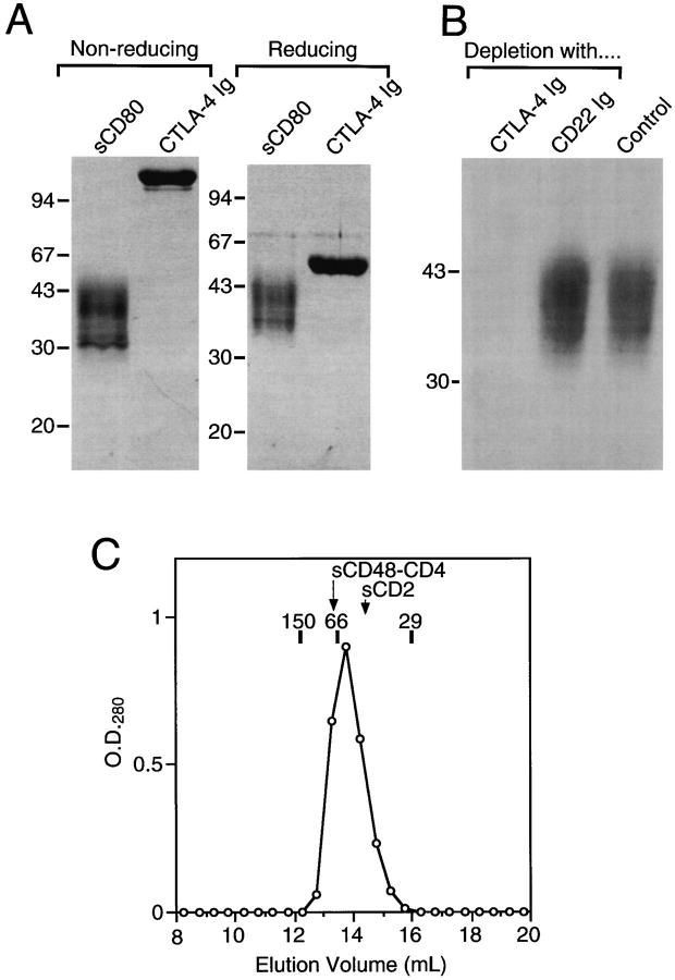
Figure 1.
Biochemical analysis of sCD80 and CTLA-4 Ig. (A) sCD80 and CTLA-4 Ig (2.5 μg each) were analyzed by SDS-PAGE on 10% acrylamide under nonreducing and reducing (+βME) conditions. The migration positions of the indicated (in kD) molecular mass markers are shown. (B) Measuring the binding activity of sCD80. Protein A–sepharose beads coated with CTLA-4 Ig or CD22 Ig were incubated with soluble sCD80, pelleted, and the supernatant analyzed for the presence of sCD80 by reducing SDS-PAGE on 12% acrylamide, together with sCD80 not exposed to beads (Control). (C) Analysis of sCD80 by size-exclusion chromatography. sCD80 (2.1 mg in 0.5 ml) was run on a SUPERDEX S200 HR10/30 column (Pharmacia) at 0.5 ml/min. The calibration markers shown (Sigma) were alcohol dehydrogenase (M r 150,000), BSA (M r 66,000), and carbonic anhydrase (M r 29,000). sCD48–CD4 and sCD2 are described in the text.

