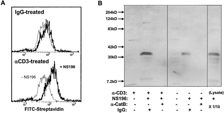
Figure 6.
Surface cathepsin B on TCR-activated T cells is active and the target of NS-196. (A) Detection of biotinylated NS-196 on the CTL surface after degranulation. Flow cytometry of CD8+ cloned human CTL RS-56 after culture on wells coated with anti-CD3 or isotope control for 2 h, followed by treatment with or without 1 μM NS-196, which was detected by FITC-streptavidin. (B) Identification of cathepsin B as the molecular target of NS-196. CTL clone RS-56 was incubated for 2 h on anti-CD3– or IgG-coated wells, followed by incubation with or without 0.1 μM NS-196. Cells were lysed with Triton X-100 and immunodepleted with beads containing anticathepsin B antibody or control rabbit IgG. The remaining lysate was run on a 12% nonreduced SDS gel, blotted onto nitrocellulose, probed with Streptavidin-HRP, and developed using ECL. The right lane shows the biotinylation pattern when the whole CTL lysate was labeled with 0.1 μM NS-196 and run directly (1/10 of the cell-equivalent input of other lanes).

