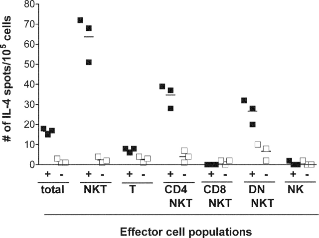
Figure 3.
GD3-reactive cells are CD4+8− NKT cells and CD4−8− NKTs. Mice were immunized with 2 × 10⁶ SK-MEL-28 cells mixed with CFA on day 0, and boosted with 2 × 10⁶ SK-MEL-28 cells mixed with IFA on day 7. Splenocytes pooled from six to eight immunized mice were separated into subpopulations based on their surface expression of NK1.1, CD3, CD4, and CD8. Total cells (unseparated splenocytes), total NKT cells (CD3+ NK1.1+), total conventional T cells (CD3+ NK1.1−), CD4+ NKT cells (CD4+ CD8− CD3+ NK1.1+), CD8+ NKT cells (CD4− CD8+ CD3+ NK1.1+), DN NKT cells (CD4− CD8− CD3+ NK1.1+), and classic NK cells (CD3− NK1.1+) populations were stimulated with irradiated syngeneic APCs in the presence (+ columns, closed squares) or absence (− columns, open squares) of GD3. IL-4 production by each cell population was detected by ELISPOT assay. Horizontal lines indicate mean values.

