
Figure 2.
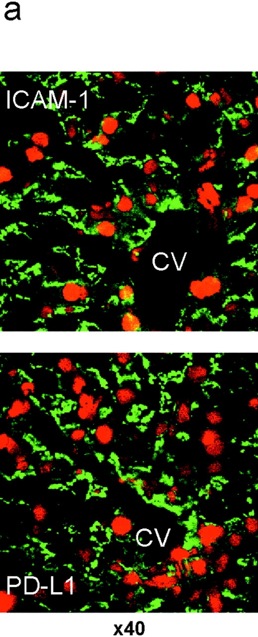
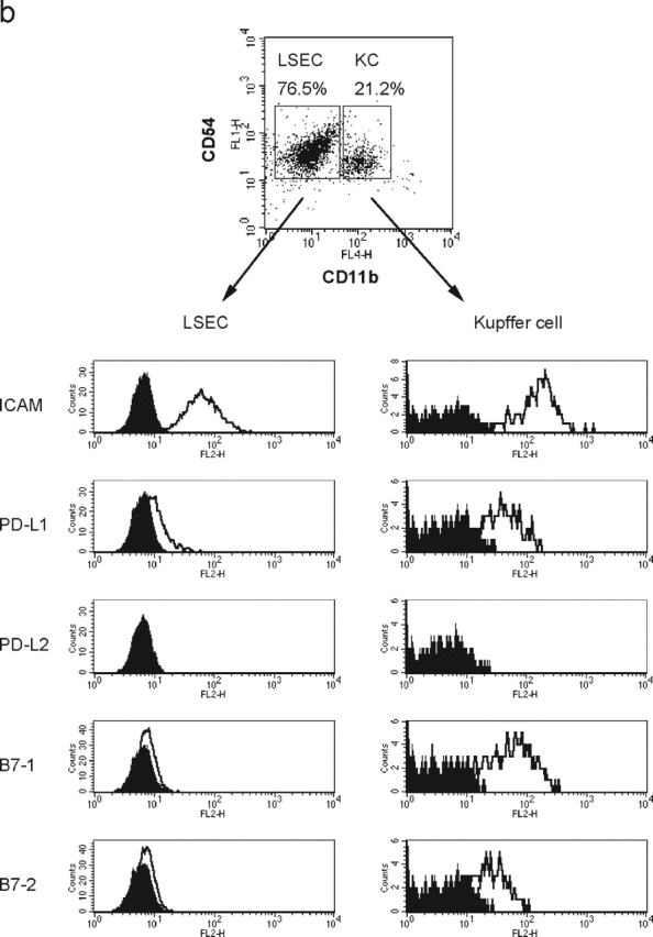
PD-L1 expression on liver nonparenchymal cells. (a) Cryosections of murine liver were stained with anti-ICAM-1 (top, green) or anti-PD-L1 (bottom, green). Nuclei were counterstained with propidium iodide (red). CV, central vein. Original magnification, ×40. (b) Surface phenotype of Kupffer cells and LSECs. LNPCs were isolated from murine livers and stained with anti-CD54 (ICAM-1)-FITC and anti-CD11b-APC, in combination with biotinylated mAb for ICAM-1, PD-L1, B7–1, or B7–2, followed by streptavidin-PE. Kupffer cells and LSECs were gated as CD54+CD11bhigh and CD54+CD11blow cells, respectively.