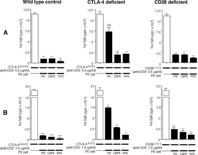
Figure 8.
Capacity of ocular PE to suppress activation of T cells from mice with disrupted CD28 and CTLA-4 (CD152) genes. PE were cultured from iris, ciliary body, and retina of C57BL/6 mice and used as substrates for purified T cells prepared from the spleens of 22-d-old wild-type mice, 22-d-old CTLA-4−/− mice, and 6-wk-old CD28−/− mice. The T cells were stimulated with anti-CD3 antibodies at concentrations of (A) 0.5 and (B) 1.0 μg/ml. Cultures were terminated at 72 h and [3H]thymidine incorporation was assessed. Mean counts per minute for triplicate cultures are presented ± SEM. Differences of means of T cell proliferation in the presence or absence of PE cells are compared. *, P < 0.05; **, P < 0.005; ***, P < 0.0005; NS, not significant.

