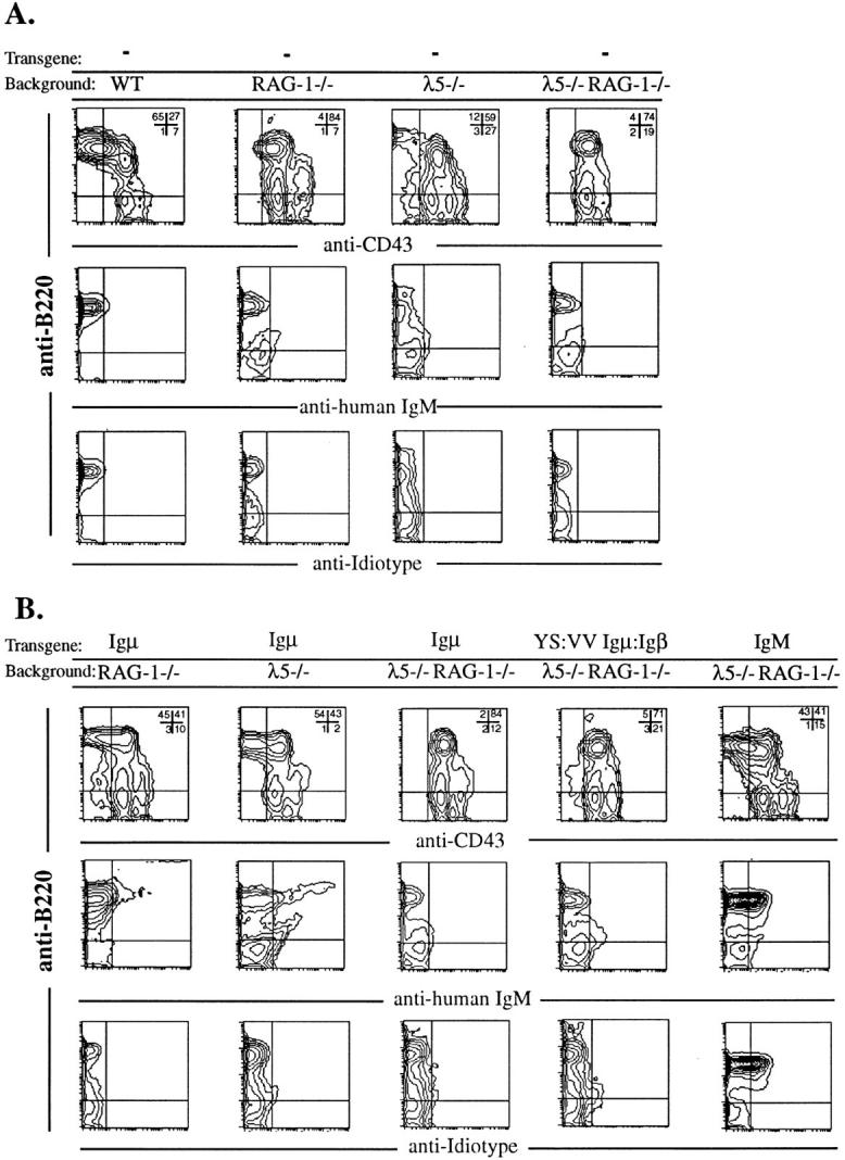
Figure 1.

Bone marrow cells from 6-8-wk old mice were analyzed by staining with PE– anti-B220, FITC–anti-CD43, biotin-anti–human IgM and biotinanti–idiotype (54.1) antibodies. The lymphocyte population was gated according to standard forward and size-scatter values. For the PE-anti-B220/FITC–antiCD43 profiles, the numbers on the upper left corner represent the percentages of gated lymphocytes in each quadrant. (A) FACS® profiles from all the control animals. (B) B cell development in the bone marrow of transgenic mice deficient for RAG-1 and λ5.
