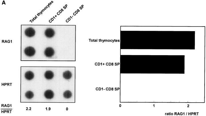
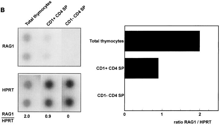
Figure 2.
RAG-1 expression of total unseparated thymocytes and CD1a− and CD1a+ CD8 (A) and CD4 SP (B) postnatal thymocytes. Thymocytes were depleted with magnetic beads for >97% of CD4 or CD8 positive cells. CD4− cells were labeled with CD8 FITC, CD1a PE, and CD3 TRC and sorted from CD1a+ and CD1a− CD3+CD8+ SP cells (A). Likewise, CD1a+ and CD1a− CD4+ SP cells were sorted from CD8− cells stained with CD4 FITC, CD1a PE, and CD3 TRC (B).


