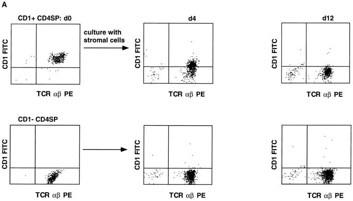
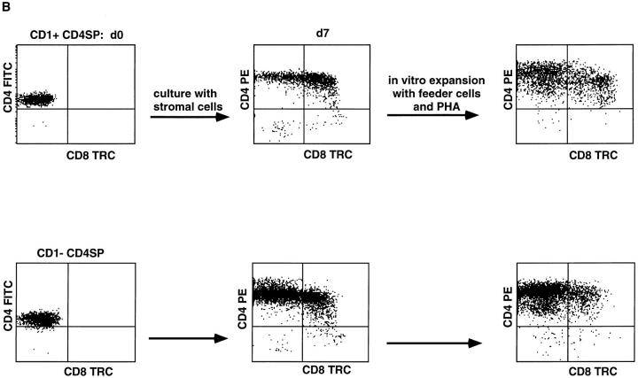
Figure 6.
CD1a+CD4+ SP thymocytes differentiate into CD1a− cells upon coculture with thymic stromal cells (A), and part of these cells upregulate CD8 expression (B). Thymocytes were labeled with CD1a FITC, CD4 PE, and CD8 TRC. CD1a+ and CD1a− CD4high SP cells were sorted and part of the cells were used to check CD3 expression by staining with CD3 TRC (all CD1a− and >99% of CD1+ CD4 SP thymocytes were CD3+). In experiment A, 2 × 10⁵ CD1a+CD4 SP (>98% purified) and 2 × 10⁵ CD1a−CD4 SP cells were cultured on a monolayer of human thymic stromal cells. After 4 and 12 d, cells were tested for CD1a expression. The cell numbers of wells started with CD1a+ and CD1a−CD4 SP cells were both reduced to 8 × 10⁴ after 4 d, whereas at day 12, 2.5 × 10⁴, and 7.0 × 10⁴ (CD1a−) cells were recovered from the cultures started with CD1a+ and CD1a−CD4 SP thymocytes, respectively. In experiment B, sorted CD1a+ and CD1a− CD4 SP thymocytes were cultured for 7 d on a monolayer of thymic stromal cells, assayed for CD4 and CD8 expression, and expanded in vitro with feeder cells, PHA, and IL-2. Expanded cells were also analysed for CD4 against CD8 expression.


