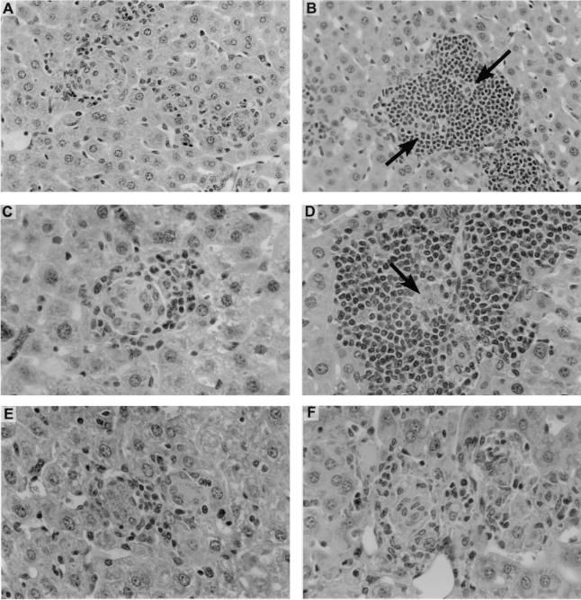
FIG. 6.
Liver histologic responses to anti-CD40 and anti-CTLA-4 MAbs in infected wild-type BALB/c mice. Mice were injected on day 12 with 0.25 mg of rat IgG or anti-CD40 or 0.5 mg of hamster IgG or anti-CTLA-4, and sections were prepared on day 21. (A and C) Infected foci in rat IgG-treated mice show early, developing granulomas. (B and D) In contrast, in anti-CD40-treated mice, parasitized foci are encased by large accumulations of mononuclear cells (arrows). (E and F) Developing granulomas in hamster IgG-treated liver (E) versus more-mature-appearing granulomas in anti-CTLA-4-injected animal (F). Panels A and B, magnification of ×180; panels C to F, magnification of ×284.

